Cave-Dwelling Blogger Hasn’t Spent Money in 9 Years
Posted: October 21, 2009 Filed under: Food and it's Impact on Our Health Leave a commentOctober 6, 2009 · Print This Article

Could you go nine years without spending a penny? It sounds pretty much impossible – how would you feed yourself, keep yourself safe from the elements? What about clothing and medicine?
Daniel Suelo consciously removed himself from the consumer lifestyle nearly a decade ago and hasn’t looked back. He lives in a cave in Utah and fishes, forages, dumpster dives and sometimes hunts for his food – and writes all about it on his website and blog from a nearby public library.
From MatadorChange, via Treehugger:
While in Ecuador on a Peace Corps mission, he witnessed a rural community acquire increased monetary wealth through farming and shift their traditional lifestyle towards a diet of unhealthy, processed food and a newfound addiction to television.
The experience led Suelo on a spiritual quest that realized itself in India, where he was particularly moved by the Sadhus, wandering monks who renounce all money and possessions. He made the conscious decision to return home, quit his job, and carve out a life without money.
As he put it, “I simply got tired of being unreal. Money is one of those intriguing things that seem real and functional because two or more people believe it is real and functional.”
Essentially an extreme freegan, Suelo receives no government assistance and does not panhandle. He lives off the excess of American society, though the kindness of strangers helps a lot when he needs a ride, and he does use taxpayer-supported public libraries.
As Treehugger points out, Suelo probably has the lowest carbon footprint of any blogger in the world. Read more about his lifestyle and how he makes it work at MatadorChange and Suelo’s own website, Living Without Money.
Imitation Foods …I call ‘em “Products”…
Posted: October 7, 2009 Filed under: Food and it's Impact on Our Health, In The Kitchen with Millie- How To's Leave a commentMany of ya”ll may not have been born by 1969. Those of us who were adults at that time know the extent to which the "new foods" really are imitation foods even though they are not labeled as such.
Crisco- a crappy, dangerous version of lard.
Margarine- A mix of vegetable oils chemically hardened to make it seem like butter..kinda.
Bouillon cubes- nothing like real stocks. Yucky and salty, and they sure don’t have that same mouth feel..mmmm.
Soy- one of the most dangerous foods; highly processed, too many dangers to go into here.

And all the other hellish stuff that man has created and people have actually learned to like..; shake and bake chicken, TV dinners, instant mashed potatoes, Kool-aid, Hawaiian punch…eeeoooowww. I learned to cook at a young age because my mom couldn’t…and I love great food, like at my grandmothers house..it was filling, healthy…at my parents I was always hungry. I whined to get apple juice instead of Kool-aid, honey instead of sugar, butter instead of margarine, Roman Meal bread instead of all the white stuff. Turns out that traditional is best..that’s why it’s a tradition. Pretty basic.
And it turns out that yes, those kitchen arts that are almost lost, stock making, canning, rendering fats, soap making…doing it from scratch is best, healthiest and saves the environment…and us. And my kids have made fun of me for loving to do things from scratch..but I love it all, paper-making, distilling flowers, making my own skin care products, using a clothesline, sewing…just has always made sense to me…and is immensely satisfying.
My next class is on Traditional Arts; Eat a Traditional Diet, How and Why, Stock and Sauce Making, Yogurt making, The Right Fats and how to use them.
A Reader Comments of the Dangers of a Vegetarian Diet
Posted: October 7, 2009 Filed under: Food and it's Impact on Our Health Leave a commentI received this comment on my blog;
 “Everyone has they’re opinion on what is right or wrong to eat? "God" speaks in the bible about what is clean and unclean to eat of that of which was provided to us. And also, the grains,fruits,etc.. Anything beyond that would be bad for you. So, it’s not what we eat or don’t eat that is detrimental or not to our health, but the man-made additives that are put into making the foods we eat so it will taste better. I do not condone the slaughter industry and the way they produce meat, however, I am not a vegetarian, but I do try to stay more vegan than meat. I’m still learning how to balance what is better, in a long run,for my health, for our bodies are not our own”.
I replied;
 While I do appreciate your opinion and input, but it just so happens that humans get very sick on just eating grains and vegetables. That’s science, not religion. I have my own "belief" system when it comes to God, but I study science and nutrition. If God had not provided meat for us to eat, and the knowledge to prepare it, the human race would not have evolved this far. Since we need meat and fats to be healthy, and God has instructed us to eat well and treat out body as a temple…so we can go forth and do his work, it cannot be wrong morally for us to eat meat.
I speak from the perspective of a recovering vegetarian. I followed that way of eating for almost 25 years…because we had no clean meat to eat! I had problems with it, it didn’t give me the right fats, I developed an allergy to EVERY SINGLE PROTEIN SOURCE associated with a vegetarian diet; SOY (after eating it for 15 years it made me start going into anaphylactic shock), DAIRY (I became seriously allergic to all dairy), NUTS (same reaction as to soy, after eating them all my life). I also became highly sensitized to grains. I got well when I stopped eating these very common allergens. Meats, fats and meats broths have enabled me to repair my immune system. I now eat a diet that man has been evolving on for thousands of years; meat, meat stocks, vegetables and fruits. Remember we have only been eating more grains after we stopped living as hunter-gatherers and began living in cities, somewhere around 1000 to 1100 AD>
Even though Kosher meat is killed kindly, it is not organic. Grass-fed gives me meat and fats that are clean; organic, full of life preserving, immune system building Vitamins A,D and E, and the right composition of fats.
I totally agree with the part of your statement about the additives and chemicals doing us harm. It is PART of the picture…but the most crucial nutrients that Americans are deficient in (calcium, Vitamin A and D, saturated fats) are only available in meat and fats from animals that have been raised in the sun!
So hang on to the limited diets that you are eating in the name of “a belief system”, but know that you will develop health problems and deficiencies as you age… Living on mostly carbs (grains and vegetables) will lead to obesity, a breakdown in your immune system, delicate skin that is prone to dryness and skin cancer, depression for lack of the right saturated fats and lack of energy and endurance.
Please read- The Ethics of Eating Meat by Charles Eisenstein
Glycemic Index on my Website
Posted: October 5, 2009 Filed under: Food and it's Impact on Our Health 3 CommentsI put a glycemic index up on my site for ya’ll to work off of. Many of the sites on line has a bazillion things to wade through because they list everything..all the breads, cereals, junk foods, sugar, candies…..stuff I don’t eat, and neither should you. So just follow the percentages; get 50% of your calories from fat, 30% from eggs, grass fed meats, and 20% from low glycemic vegetables. Never go above 50 on the glycemic index…or not very often. I always have fruit at breakfast with my fats and proteins, and veggies with lunch and dinner.
Optimum Nutrition Glycemic Index
The Dirty Secret about Agave
Posted: October 5, 2009 Filed under: Food and it's Impact on Our Health 5 CommentsThe process by which agave glucose and inulin are converted into “nectar” is similar to the process by which corn starch is converted into HFCS.
 The skinny on natural sugar alternatives is that they are a big, fat business opportunity and therefore worth a closer look. You know that our species is genetically programmed to eat sweets until we pretty much explode. Savvy consumers are constantly trying to avoid not only the dizzying amount of sugar and high-fructose corn syrup in our food supply, but also spooky artificial sweeteners, most of which were discovered by accident by people wearing safety goggles and lab coats. This leaves us to appease our sweet tooth with natural sweeteners, a few of which are having a “moment.” But are they—like Ponzi schemes, unnaturally muscular athletes, and Tequila-born love affairs—just too good to be true?
Ina word…YES!
In spite of manufacturers’ claims, agave “nectar” is not made from the sap of the yucca or agave plant but from the starch of the giant pineapple-like, root bulb. The principal constituent of the agave root is starch, similar to the starch in corn or rice, and a complex carbohydrate called inulin, which is made up of chains of fructose molecules. Technically a highly indigestible fiber, inulin, which does not taste sweet, comprises about half of the carbohydrate content of agave. 34
The process by which agave glucose and inulin are converted into “nectar” is similar to the process by which corn starch is converted into HFCS. 35
The agave starch is subject to an enzymatic and chemical process that converts the starch into a fructose-rich syrup—anywhere from 70 percent fructose and higher according to the agave nectar chemical profiles posted on agave nectar websites.
36
(One agave manufacturer claims that his product is made with “natural” enzymes.) That’s right, the refined fructose in agave nectar is much more concentrated than the fructose in HFCS. For comparison, the high fructose corn syrup used in sodas is 55 percent refined fructose. (A natural agave product does exist in Mexico, a molasses type of syrup from concentrated plant nectar, but availability is limited and it is expensive to produce.)
According to Bianchi, agave “nectar” and HFCS “are indeed made the same way, using a highly chemical process with genetically modified enzymes. They are also using caustic acids, clarifiers, filtration chemicals and so forth in the conversion of agave starches.” The result is a high level of highly refined fructose in the remaining syrup, along with some remaining inulin.
In a confidential FDA letter, Dr. Martin Stutsman of the Food and Drug Administration’s Offce of Labeling Enforcement, explains the FDA’s food labeling laws related to agave nectar: “Corn syrup treated with enzymes to enhance the fructose levels is to be labeled ‘High Fructose Corn Syrup.’” According to Mr. Stutsman, agave requires the label “hydrolyzed inulin syrup.”37
Corn Syrup.’” According to Mr. Stutsman, agave requires the label “hydrolyzed inulin syrup.”37 Even though, like corn, agave is a starch and fber food processed with enzymes, it does not require the label “High Fructose Agave Syrup.” Agave “nectar” is a misnomer; at the very least, it should be labeled “agave syrup.”
Agave syrup comes in two colors: clear or light, and amber. What is this difference? Mr. Bianchi explains: “Due to poor quality control in the agave processing plants in Mexico, sometimes the fructose gets burned after being heated above 140 degrees Fahrenheit, thus creating a darker, or amber color.” However, the labels create the impression of an artisan product—like light or amber beer.
THE SAPONIN PROBLEM
Yucca species are known to contain large quantities of saponins. The industry describes saponins in agave syrup as beneficial: “Agave’s rich density of saponins increases hydration as the soapy, surfactant nature of saponins change the wetting angle of water it contacts. This eases and accelerates cellular water uptake, especially when used with a high-quality salt.”38
However, the truth is that the saponins found in many varieties of agave plants are toxic steroid derivatives, capable of disrupting red blood cells and producing diarrhea and vomiting,39 Agave should be avoided during pregnancy or breastfeeding because they might cause or contribute to miscarriage by stimulating blood flow to the uterus.40 At the very least, agave products should carry a warning label indicating that the product may cause a miscarriage.
JUST SAY NO TO AGAVE
Since the FDA makes no effort to enforce food-labeling laws, consumers cannot be certain that what they are eating is what the label says it is. New sweeteners like agave syrup were introduced into the market to make a profit, not to make consumers healthy. Clever marketing has led mane consumers to believe that the high level of fructose in agave syrup makes it a safe and a natural sweetener. Agave syrup labels do not conform to FDA labeling requirements, thus deepening the false illusion of an unprocessed product. As we have demonstrated here, if a sweetener contains manufactured fructose, it is neither safe, nor natural, especially at levels up to 70 percent.
Agave syrup is a manmade sweetener which has been through a complicated chemical refining process of enzymatic digestion that converts the starch and fiber into the unbound, manmade chemical fructose. While high fructose agave syrup won’t spike your blood glucose levels, the fructose in it may cause mineral depletion, liver inflammation, hardening of the arteries, insulin resistance leading to diabetes, high blood pres-sure, cardiovascular disease and obesity.
If you want something sweet, eat a piece of fruit, not a candy bar labeled as a “health food.”
If you want to create something sweet, use sweeteners that are known to be safer. For uncooked dishes, unheated raw honey or dates work well. For cooked dishes or sweet drinks, a good organic maple syrup, or even freshly juiced apple
juice or orange juice can provide delicious and relatively safe sweetness; dehydrated cane sugar juice or maple sugar may be used in moderation in cookies and desserts that contain nutritious ingredients and good fats such as butter, egg yolks and nuts.
However, to be healthy, we cannot eat sugar all day, no matter how natural the form. One should limit total sweetener consumption to less han fve percent of daily calories. For a diet of 2500 calories per day, that’s less than three tablespoons of honey, maple syrup or dehydrated cane sugar juice, or several pieces of fruit. And many people do best by avoiding sweeteners completely.
The lack of standards in the health food world comes as depressing news; but let this news encourage you to consume more pure and unrefined foods and sweetener sources. Good health depends on wise food choices, and wise food choices depend on constant vigilance.
More on the Agave Industry
In the year 2000, with warrants in hand, federal agents from the Office of Criminal Investigations of the Food and Drug Administration (FDA) came banging on the door of North America’s largest agave nectar distributor, Western Commerce Corporation in California. In an extremely rare case of the FDA protecting consumer interests (rather than supporting big business, while shutting down legitimate and health consciousness competition), they discovered that Western Commerce Corporation was adulterating their agave syrup with high fructose corn syrup (to lower the cost even more and increase profit margins). While the federal agents confiscated material in the warehouse, the owners of Western Commerce Corporation were nowhere to be found. Those who ran the company fled the country with millions of dollars in assets to avoid criminal prosecution.
This adulterated agave syrup (refined fructose) was also labeled as certified organic to fool consumers into thinking they were getting a pure product. This shows you how unverified organic labels are used in the USA. Today, high fructose agave syrup is made primarily by two companies: Nekutli, and IIDEA. A third agave marketer, by the name of Volcanic, has a suspicious claim on their website. “If your agave comes from one of the other two companies in Mexico, something has been added.” They are referring to Nekutli and IIDEA. Their claim is based upon an analysis, which they say shows that Volcanic’s agave nectar has a lower level of refined fructose.
When Western Commerce Corporation was shut down, the large retail establishments in the food industry stayed away from agave syrups. They knew better than to risk lawsuits and consumer fraud. “They understood that agave was criminally mislabeled per the U.S. Code of Federal Regulation labeling laws, with an untried sweetener, new to the market, that contained saponins, and was not clearly approved as safe for use,” explains Mr. Bianchi. For many years following this bust, the supermarket and health food store industry avoided using agave.
But recently, some sellers in the agave syrup field, once quiet, have begun sneaking back into the food and beverage chains. And retail food giants like Whole Foods, Wegman’s, Trader Joe’s and Kroger, who should know better, and who should know the food labeling laws and requirements, still have no hesitation in selling the toxic, unapproved and mislabeled refined fructose agave syrup, as well as products containing it.
Source: Russ Bianchi and the Weston Price Foundation
For References click HERE
24 Hours into the Beef Stock Making…
Posted: October 5, 2009 Filed under: Food and it's Impact on Our Health, In The Kitchen with Millie- How To's Leave a commentThe huge bones are simmering on the stove..I felt like a cave woman in my kitchen last night…browning huge beef shank bones in my Dutch oven, in luscious coconut oil. So tonight it has been sloowly bubbling for 24 hours..I added carrots, onions, celery..all organic, with bay leaves, crushed peppercorns…Celtic salt.
And as I just tasted it for the first time…I realized that I have never tasted a true beef stock before…the mouth feel is nothing like the insipid, salty stuff I have had. The grass fed beef is heavenly to cook with…amazingly nutritious. The gelatin is developing, it’s silky texture is amazingly satisfying.
Another 24 hours to go…the house smells amazing.
I’m making beet salad from beets I grew, then roasted with the beef bones in the oven last night. I added dill and rice vinegar, light salt. Mmmm
While it’s bubbling….I’m making a flourless chocolate cake. Here’s the recipe;
Flourless Chocolate Cake Topped with Raspberries

- 12 oz semisweet chocolate chips
- 1/2 cup butter
- 1/4 cup sugar
- 1/4 cup water
- 1 teaspoon instant coffee granules
- 3 eggs
Preheat the oven to 425 F.
Butter the bottom and sides of a 9 inch pie plate.
Place a 12 inch square sheet of waxed paper in the plate and butter the waxed paper.
In a medium sized saucepan, combine the chocolate chips, 1/2 cup butter, sugar, water, and coffee granules. Heat over medium heat for 2 to 3 minutes, until the chocolate and butter are melted, stirring constantly.
Remove from the heat and stir in the eggs until the mixture is smooth. Pour into the waxed paper-lined pie plate. Bake for 10 minutes; the cake will not be completely set in the middle. Cool, cover loosely, then chill for 6 to 8 hours, or overnight.
When ready to serve, remove the cake from the refrigerator and allow to sit for 10 minutes. Invert the cake onto a large flat serving dish and remove the waxed paper.
Top with raspberries across the whole top, begin in the middle and work out…letting the raspberry tips straight up.
![]()
![]()
![]()
Virgin Coconut Oil from Tropical Traditions…
Posted: October 4, 2009 Filed under: Food and it's Impact on Our Health Leave a comment Close friends of mine began nagging me a few years ago to check out the company that they were getting coconut oil from, Tropical Traditions. They said the thier coconut oil was the best they’d ever had; unprocessed, made by hand, organic… I had only a few years before began researching saturated fats and the role they play in the body, and eating coconut oil…and was pleased with the organic coconut oil I was already eating. Plus, money was tight and I saw no reason to change to a more expensive product…until I went on the site and read about this wonderful product, and began looking into the differences in virgin oils compared to processed oils.
About 3 weeks ago my friends told me they were having an awesome sale, half off on coconut cream, so I ordered some of thier virgin oil. They sent me a book on the company, how the coconut oil was handmade and the health benefits of unprocessed virgin oil.
First of all, the service was wonderful, customer service was excellent, I got my order quickly…the coconut oil and the cream were in glass jars, not plastic. The packaging was all cardboard, no Styrofoam, no plastic.
I love it. I like the taste, the coconut cream is amazing…I made Coconut Fudge Sauce and it is to die for! The cream is coconut concentrate, I use it just like I would heavy cream in a recipe.
The oil I cook with, bake with, use on my hair, my face, my legs after shaving… Coconut cream has always been my favorite oil, mixed with olive oil..to make bar soap or shampoo.
They also carry coconut; shredded, chips, flour (WAY lower on the glycemic index than other flours, a dream to bake with and gluten free!), They carry organic unrefined palm oil, sesame, extra virgin olive oil, soaps, grass fed meat, organic foods.
Here is what info from thier website;
Independent laboratory analysis shows this to be one of the highest quality coconut oils on the market, having the highest levels of antioxidants. This enhanced Virgin Coconut Oil is now in the US market under the Tropical Traditions Gold Label brand. It meets our strictest standards to earn this designation. Today when you buy Tropical Traditions Gold Label Virgin Coconut Oil, you are buying the highest quality coconut oil we have to offer, and it is still made by hand and benefiting families in the rural areas of the Philippines where the coconuts grow.

Is Virgin Coconut Oil the healthiest oil on earth?
Read the research here!
Read what Weston Price has to say about Coconut Oil.
Characteristics of Our Gold Label Virgin Coconut Oil
This Tropical Traditions Gold Label Virgin Coconut Oil is a truly unrefined coconut oil. This coconut oil is made on Mt. Banahaw and surrounding areas from organic coconuts. Coconuts are used fresh (within 24-48 hours of harvest) from small family farms on Mt. Banahaw and other rural places in Quezon Province, the coconut capital of the Philippines. Only the highest quality coconuts are hand-picked from each harvest. The volcanic soil of Mt. Banahaw makes these organic coconuts some of the most nutritionally rich coconuts in the world! Testing done in independent laboratories reveals higher levels of phenolic antioxidants than other coconut oils. The fresh coconut meat is shredded (wet milled), and then cold-pressed using the water from inside the coconuts to make coconut milk. The milk is then allowed to sit for about half a day, while the oil naturally separates from the heavier water. The oil is then filtered from the curds (coconut solids). No chemical or high-heat treatment is used, and this oil contains no trans fatty acids. We do NOT mass produce this oil. It is made by families who are coconut farmers using old-fashioned traditional methods that have been used in the Philippines for hundreds of years. Our coconut trees and family producers are certified organic according to strict USDA standards.
Tropical Traditions also has a Referral Program, so if you place an order with them as a first-time customer, please select “Referred by a friend” and in the box that says “How did you hear of us?” enter my sponsor ID number… 5541380. By telling Tropical Traditions that I referred you, you will receive a complimentary copy of the book Virgin Coconut Oil: How it has changed people’s lives and how it can change yours! by Brian and Marianita Shilhavy with your first order! This book is filled with testimonies and research showing how healthy coconut oil is, and it also includes over 85 recipes showing how one can incorporate coconut into their diet.
I am thrilled to have found this product and recommend it highly.
The Beef Bones Adventure…
Posted: October 4, 2009 Filed under: Food and it's Impact on Our Health, In The Kitchen with Millie- How To's 1 Comment I have been happily making chicken stock for quite some time now.. About a year and a half ago, while working at Native Sun, I kept bugging the employees in the meat department about finding me a case of grass fed beef shank bones. Months this went on, to no avail. They said they couldn’t get an answer…then they said no.
I could find them on the internet, but I’ll go to extremes to keep from having something shipped to me..for environmental reasons mainly..but also wanting really fresh bones. The local co-ops want 35. to 50. to join.. What to do? I finally sucked up and went to Whole Foods…whom I certainly don’t trust for lots of reasons, preferring to shop local, not chains..
But a friend called and said she had asked them about the bones and they had them…so I got up the next morning and schlepped all the way out there. and was told they didn’t have them. They apologized and gave me the meat managers card, who would be in the next afternoon. So I waited til then and called, spoke to him and made arrangements to drive out the next afternoon to get them..he would hold them for me.
He didn’t…or rather when I got there, again, the next afternoon in 5 o’clock traffic…he wasn’t there. Seems he had been taken to the hospital for diabetes. Why don’t health food stores teach people how to eat? Classes on nutrition? Anyway, I whine, they look all over, can’t find them. They talked to the store manager, who had them cut 4 the meat off of 3 shanks…a 40 minutes wait…but t5hey did it. While hanging out at the meat counter, I noticed that bison was on sale for 3.99 a POUND. Wow… bought three pounds… Finally heaved the bones out to the car, with help…and at a great price.
The burger I had that night was the best bison burger I’ve ever had, very fresh, perfect. Wow.
And this afternoon I came home and browned, then slow roasted all those shanks and they are gently simmering for the next 2 days..mmmmmmmm.
Beef Stock

about 5 pounds beef shank bones
2 Tablespoons coconut oil
3 pounds meaty rib or neck bones
5 or more quarts cold filtered water
1/2 cup vinegar
3 onions, coarsely chopped
3 carrots, coarsely chopped
several sprigs of fresh thyme, tied together
Heat coconut oil to slightly above medium heat, place shanks bones in oil to brown. Brown on all sides, even the ends of marrow. Then place in a roasting pan, covered, and brown at 325 degrees in the oven for 1 hour.
Place the knuckle and marrow bones in a large pot with vinegar and cover with water. Let stand for one hour.
Place roasted bones the water with them knuckles and stuff. Slowly bring up to a soft simmer and simmer, covered for…the longer the better..up to 72 hours.
Pour the fat out of the roasting pan. Deglaze the roasting pan, add liquid to pot., Add additional water, if necessary, to cover the bones; but the liquid should come no higher than within one inch of the rim of the pot, as the volume expands slightly during cooking. Bring to a boil.
A large amount of scum will come to the top, and it is important to remove this with a spoon. After you have skimmed, reduce heat and add the thyme and crushed peppercorns.
Simmer stock for at least 12 and as long as 72 hours. You will now have a pot of rather repulsive-looking brown liquid containing globs of gelatinous and fatty material. It doesn’t even smell particularly good. But don’t despair. After straining you will have a delicious and nourishing clear broth that forms the basis for many other recipes.
After you have made the stock and reduced it as much as you want, place the pot in the refrigerator and let it cool overnight. All the fat will rise to the top. Boil that fat with twice the amount of water as fat, let it solidify again, and THEN store it in the fridge or freezer. This removes a good amount of the meaty taste, and whatever sediment is in the fat will sink to the bottom of the pot. Use it like you would schmaltz or any other solid fat — especially for frying or in pie crusts!
You can do this with bacon grease, too, although I don’t know why you would want to get RID of bacon flavor!! 🙂
High Cholesterol = Heart Disease — Myth or Truth?
Posted: October 1, 2009 Filed under: Food and it's Impact on Our Health 5 CommentsThe Response-to-Injury Rabbit Never Developed Atherosclerosis — Why Not?

by Chris Masterjohn
From Cholesterol and Health.com
The pop science version of cholesterol goes something like this: when you eat fatty foods, especially foods rich in animal fat, the saturated fat and cholesterol in these foods wind up in your blood and stick to your arteries. Since saturated fats are solid outside your body, they will be solid inside your body too — depsite the 30-degree increase in average temperature. Arteries are much like pipes. When they get caked up with grease, blood flow is impaired, and a heart attack ensues.
None of the prominent scientists who promoted the idea that cholesterol is a critical factor in the development of heart disease ever believed anything remotely resembling this nonsense. From the beginning, they recognized that atherosclerotic plaque accumulates behind the layer of the artery in contact with the blood, called the endothelium, and that the cholesterol and fat within it is engulfed in white blood cells.
The theory these scientists promoted looked something like this: when the cholesterol level in the blood increases, it penetrates the arterial wall and gets stuck; white blood cells circulating in the blood then enter the arterial wall and gobble up the cholesterol; the accumulation of lipid-loaded white blood cells causes local injury, leading to cell death, calcification, and the development of a collagen-laden "fibrous cap" over the atherosclerotic lesion. When the cap ruptures, the blood clots, blocking the artery and causing a heart attack. This is called the lipid hypothesis.
But is this true? Books and web sites devoted to debunking this theory have come out of the woodwork over the last decade; books defending it have followed suit. Consider the following titles to see just how controversial the idea really is:
- The Cholesterol Myths: Exposing the Fallacy That Saturated Fat and Cholesterol Cause Heart Disease by Uffe Ravnskov, MD, PhD (2000).
- The Great Cholesterol Con: Why everything you’ve been told about cholesterol, diet, and heart disease is wrong! by Anthony Colpo (2006).
- The Cholesterol Wars: The Skeptics vs. the Preponderance of the Evidence by Daniel Steinberg, MD, PhD (2007).
- The Great Cholesterol Con: The Truth About What Really Causes Heart Disease and How to Avoid It by Malcolm Kendrick, MD (2007).
So is the theory that cholesterol causes heart disease just a myth? Or are the skeptics truly waging a war against the preponderance of the evidence?
The Cholesterol Debate — What Causes Atherosclerosis?
The truth is that each of these authors makes important points. Were there never any good evidence that cholesterol was involved in heart disease, there would be no National Cholesterol Education Program, no statin empire, and Daniel Steinberg could never have written a book plus over 200 scientific papers on the subject. On the other hand, were there never anything seriously wrong with the mainstream dogma on the issue, Ravnskov, Colpo, Kendrick, and many other authors could never have built their careers around pointing out the gaping holes in the theory.
There is no one cause of "heart disease." "Heart disease" is a heterogeneous compliation of diseases of the heart and blood vessels with many different causes. Some of these include disturbances of the rhythm of the heart, calcification of the middle portion of the blood vessels and calcification of the heart valves, and congestive heart failure. The question I address in this article is whether and in what sense cholesterol is involved in atherosclerosis, the development of fatty and calcified plaques in isolated, raised lesions, which can cause heart attacks by rupturing, clotting, and blocking arteries.
In 1933, the famous proponent of the cholesterol-fed rabbit model Nikolai Anitschkov declared that atherosclerosis had been shown to be of an "infiltrative" character rather than a "degenerative" character and was driven by lipids (fatty substances) rather than by inflammation. He did not deny inflammation was involved, but believed that it was secondary to lipid infiltration. Many opponents continue to claim that the root cause driving heart disease has nothing to do with lipids and everything to do with inflammation and that it is degenerative rather than infiltrative in character.
As we will see below, these are all correct! Atherosclerosis is largely driven by the degeneration of lipids which infiltrate the blood vessel and thereby cause inflammation. Inflammation from other sources may accelerate the process or further the degeneration of the atherosclerotic plaques once they are formed, but the initiating factor for fatty plaques appears to be the degeneration of lipids — especially the degeneration of polyunsaturated fatty acids (PUFA).
In order to begin looking at the evidence, we must go back a century in time to the cholesterol-fed rabbit. The cholesterol-fed rabbit model came on the heels of extensive investigations into what would later be termed the "response-to-injury hypothesis."
The Response-to-Injury Rabbit Model
Around the turn of the twentieth century, research into the cause or causes of heart disease was in full throttle. A 1933 compilation edited by E.V. Cowdry entitled Arteriosclerosis: A Survey of the Problem (New York: Macmillan) contained twenty reviews of investigations into the matter, including statistical relationships, the distribution of the disease in wild animals, the distribution in humans according to race and climate, nutritional influences, the physical and chemical nature of the changes that occur in atherosclerotic tissues, and experimental models of the disease.
Nikolai Anitschkov, who developed the cholesterol-fed rabbit model, wrote the 50-page review of experimental animal models.1 Much of this research was published in German, so Anitschkov’s review is an invaluable resource.
According to Anitschkov, early ideas about the origin of arteriosclerosis — a general term for hardening and damage to the arteries, of which atherosclerosis is a specific type — saw the diseases as a response to injury. The injury was primarily seen as either a mechanical or a toxic factor, and was sometimes believed to be injury to the nerves rather than injury to the blood vessels. Researchers carried out a multitude of experiments on rabbits and other animals, including the following:
- Mechanical damage to the blood vessels including ligating, pulling, pinching, and wounding them, and cauterizing them with galvanic wire or silver nitrate.
- Increasing blood pressure by constricting the blood supply through the aorta, damaging the kidneys, or hanging rabbits up by their feet.
- Severing or irritating certain nerves.
- Injecting rabbits with adrenalin.
- Injecting rabbits with a multitude of toxic factors, including digitalin, strophanthin, adonidin, ergotin, theocin, barium chloride, hydrastin, nicotine, caffeine, formalin, ergosterol, and various salts of acids and heavy metals.
- Injection of diphtheria toxin and many other bacteria cultures or bacterial byproducts.
Most of these methods caused substantial damage to the arteries and resulted in a "regenerative thickening" of one or another type. So the response-to-injury concept is quite real.
Atherosclerosis is Just One Type of Arteriosclerosis
None of these methods, however, produced anything resembling human atherosclerosis. While arteriosclerosis refers to hardening and degeneration of the arteries in general, atherosclerosis is a specific type of arteriosclerosis in which a plaque rich in lipid-loaded white blood cells, cholesterol, fatty acids, calcium, various debris — called an atheroma — invades the innermost layer of the blood vessel wall called the intima, just behind the one-cell-thick layer called the endothelium. If you are not familiar with the anatomy of a blood vessel, you can see a diagram of it here.
The research in Anitschkov’s day suggested that, while various types of arteriosclerosis occurred in humans, atherosclerosis was a much more important cause of death. Anitschkov thus concerned his research with what caused atherosclerosis.
The mechanical injuries to blood vessels or nerves produced a local repair process that involved the proliferation of cells, their congregation around the damaged area, and a resultant thickening of the vessel wall. The results were local rather than systemic, however, and never produced a lesion resembling an atheroma.
Injections of adrenalin produced much more interesting changes that were much more relevant to humans. They produced necrosis (death) of cells in the media followed by extensive calcification. A similar process was observed in some of the blood pressure experiments and in many of the experiments involving injections of metallic, bacterial, or other toxins. These changes, however, were fundamentally different from atherosclerosis, which occurs in the intima.
Medial Calcification and the Vitamin K2 Connection
That does not mean this research is irrelevant. Humans experience this type of medial calcification in diabetes, kidney disease, and aging. It appears to assault the media of arteries and the valves of the heart together. It increases arterial stiffness and decreases the artery’s ability to accomodate moderately high levels of blood pressure. One of the most important factors in this type of calcification appears to be vitamin K2.
Vitamin K-dependent proteins protect against cell death, help clear away the debris that cells leave behind when they do die, and protect against the calcification of soft tissues. In the absence of sufficient vitamin K, these proteins are deformed and fail to work properly. It appears that vitamin K2, found in animal fats and fermented foods, is far more important in this respect than vitamin K1, found in green plant foods. I have written extensively on this subject and argued that vitamin K2 is the "activator X" of Weston Price in my article, On the Trail of the Elusive X Factor: Vitamin K2 Revealed.
Despite the research in Anitschkov’s day suggesting that only atherosclerosis had major clinical importance, research in our own day shows that calcification of the media and valves is critically important to, at a minimum, the 324 million people worldwide who will be diabetic come 2025. For the US population born in 2000, the estimated lifetime risk of type 2 diabetes is one in three.2 In type 2 diabetics, medial calcification increases the risk of mortality from heart disease, stroke, and all causes. It also predicts the incidence of heart disease and stroke, including events that do not produce fatalities, and predicts the likelihood that peripheral artery disease will require limb amputation.3
So the response-to-injury hypothesis has a solid basis of evidence for arteriosclerosis of the media, and this is clinically important — but what causes atheroma, that is, the fatty plaque that causes raised lesions in the intima of the blood vessels?
To answer this question, we must look to the cholesterol-fed rabbit.
The Cholesterol-Fed Rabbit Controversy
In 1909, a researcher at the Military Medical Academy in St. Petersburg named Ignatowski produced atherosclerosis in rabbits by feeding them a diet of meat, eggs, and milk. He was pursuing a hypothesis put forward by Nobel Prize-winning microbiologist I. Metchnikov that dietary protein accelerated aging.4
In 1913, Anitschkov and his partner Chalatov were studying at the same academy and were assigned to follow up Ignatowski’s work. They progressively narrowed down the causative factor to cholesterol by feeding different foods and fractions of foods, finally producing the diease by feeding pure cholesterol dissolved in sunflower oil.4
Rabbits fed sunflower oil alone did not develop atherosclerosis. In the cholesterol-fed rabbits, however, lesions developed that exhibited a remarkable similarity to the human disease. They began as fatty streaks in the intima; circulating white blood cells then invaded the intima and engulfed the cholesterol and fat deposited there, eventually growing into large phagocytic cells that Anitschkov called xanthoma cells and we now call foam cells; eventually the developing plaque protruded into the intima in the form of a raised lesion. The lesion possessed a fatty core rich in crystalized and calcified cholesterol deposits and was covered with a fibrous cap.1
The lesions did not appear everywhere equally, but occurred in specific areas. They were most prominent in the aorta and other large arteries, especially in the areas of the artery wall that experience disturbed blood flow such as the points where the arteries branch. While they did not occur in exactly the same places as human atherosclerotic lesions, the pattern was largely similar and the underlying physiological principle dictating the location of the lesions — mainly the type of blood flow experienced by the artery wall — was the same.1
The rabbits developed cholesterol deposits all throughout their bodies, in their eyes and internal organs. Anitschkov produced a more mild form of the disease, however, by feeding the rabbits milk. In these experiments, the rabbits received a much more moderate amount of cholesterol over a much longer period of time and the resulting disease was much more focused in the arteries.1
One curious difference between rabbits and humans is that when rabbits develop atherosclerosis, their plaques never rupture and they never get heart attacks. The main determinant of plaque rupture according to the current scientific literature is the balance between collagen degradation and collagen synthesis.5 Collagen synthesis requires vitamin C. Most animals, including rabbits, make their own vitamin C, but humans do not.
Atherosclerosis itself probably diminishes the quality of life in many different ways by impeding blood flow and blood vessel function, but it clearly does not inexorably lead to heart attacks. The reason why atherosclerosis produces heart attacks in humans and not rabbits or many other animals might be that humans cannot produce their own vitamin C.
Cholesterol in the Blood, Not the Food
Anitschkov argued against calling cholesterol "the cause" of atherosclerosis, but he considered cholesterol the primary causal factor and the necessary causal factor. Mechanical injuries, adrenalin injections, and other methods used to induce various types of arteriosclerosis would accelerate the development of atheroma when they were combined with cholesterol-feeding, but they would never result in human-like atherosclerosis by themselves.
Anitschkov never concluded from his experiments that cholesterol in the diet caused atherosclerosis in humans, however. To the contrary, he wrote the following:
[I]n human atherosclerosis the conditions are different. It is quite certain that such large quantities of cholesterin are not ingested with the ordinary food. In human patients we have probably to deal with a primary disturbance of the cholesterin metabolism, which may lead to atherosclerosis even if the hypercholesterinemia is less pronounced, provided only that it is of long duration and associated with other injurious factors.
Cholesterol skeptics often argue that the rabbit is irrelevant to the human because it is an herbivore. Cholesterol-feeding has failed to produce atherosclerosis in many other species. This is true, but it misses the point. In the species where cholesterol-feeding alone does not produce atherosclerosis, the blood level of cholesterol does not rise as much as in rabbits. But in all of these species when the level of cholesterol in the blood rises high enough, atherosclerosis ensues. For example, feeding dogs cholesterol alone does not produce atherosclerosis because they turn the cholesterol into bile acids; but inhibiting thyroid hormone stops them from making this conversion, and when combined with cholesterol-feeding, it induces atherosclerosis.
As Steinberg points out, raising blood levels of cholesterol has produced atherosclerosis in baboons, cats, chickens, chimpanzees, dogs, goats, guinea pigs, hamsters, monkeys, mice, parrots, pigs, pigeons, rabbits and rats.
The role of blood cholesterol in human heart disease was supported by research showing that people with a disorder that would eventually be called familial hypercholesterolemia had dramatically increased blood levels of cholesterol and, in youth and middle age, dramatically increased relative risks of heart disease and atherosclerosis. But what caused their high cholesterol levels, and did those levels cause the atherosclerosis, and if so, did this phenomenon have any relevance to the rest of us?
And, if cholesterol was somehow the culprit in all of this, was it merely its concentration in the blood that was at play, or was something very different going on?
Lessons From Familial Hypercholesterolemia
Familial hypercholesterolemia (FH) bears a striking resemblance to the cholesterol-fed rabbit model. In mild cases, it produces earlier and more rapidly developing atherosclerosis compared to the general population. In its severe cases, it results in cholesterol deposits all throughout the body, especially in the liver, kidneys, and eyelids.4
In the mid-1970s, Brown and Goldstein discovered that FH resulted from a single genetic defect in the LDL receptor that made the cells unable to absorb LDL from the bloodstream. Steinberg argues that, since cells jealously guard their cholesterol concentrations by adjusting their synthesis of cholesterol as needed, this showed that FH patients differed from the general population in only one single way: the concentration of cholesterol in their blood.4
The finding drew several more parallels between FH and Anitschkov’s cholesterol-fed rabbits. Anitschkov argued that it was not the mere feeding of cholesterol to the rabbits that produced atherosclerosis, but the overwhelming of their capacity to use and dispose of that cholesterol. FH cells could absorb free cholesterol, but not cholesterol from LDL. Anitschkov’s rabbits developed atherosclerosis when they ate cholesterol, but not when they were injected with it — in which case it would not be packaged into lipoproteins such as LDL, which contain many other substances besides cholesterol. Looking backward, it appears that the common thread running through each model was that the level of LDL in the blood exceeded the capacity of the LDL receptors to move that LDL from the blood to the cells.
The LDL receptor highway was blocked, and the LDL traffic was jammed.
Is Steinberg correct, however, that this changes nothing but the concentration of LDL in the blood? Consider what happens in a traffic jam:
- The concentration of cars in the road increases.
- It takes you longer to get home.
When LDL can’t get from the blood into the cells, its concentration in the blood rises, but it also spends a longer amount of time in the blood. Why would that matter? This would become clear just several years later. At the end of the 1970s, the role of oxidative stress in heart disease would finally become clear.
The Role of Oxidized LDL in Heart Disease
Anitschkov believed that his research showed that atherosclerosis was of an infiltrative character rather than a degenerative character. He believed that cholesterol and other substances naturally permeated the endothelium in order to nourish the other layers of the blood vessels, and proceeded from there into the lymph fluid. When the blood level of cholesterol rose sufficiently, he argued, it entered the intima at a faster rate than it could exit and began to accumulate.
Anitschkov was correct that the disease was driven by an infiltration of lipid, and he was correct that the degeneration of the blood vessel wall was secondary to this infiltration. What he failed to realize, and could not have realized at the time, was that the entire process depended on the degeneration of the lipid.
The Discovery of Oxidized LDL
Beginning in 1979, investigators made a series of revolutionary discoveries revealing this degenerative process. When they incubated cells with LDL in the absence of other serum components, the cells underwent severe damage and began to die within 24 hours. Adding serum or HDL prevented the toxicity.4
In 1981, these researchers discovered that culturing endothelial cells with LDL caused dramatic changes to the LDL, making it denser, more electronegative, and giving it a dramatic ability to accumulate in white blood cells called macrophages. Macrophages are phagocytic, meaning they like to gobble up other things, and they are the precursors to the "foam cells" that populate atherosclerotic plaques. The researchers called this LDL "endothelial cell-modified LDL." Soon after, they discovered that the LDL was being "oxidatively modified" and that not only HDL but vitamin E (which HDL is rich in) prevented the effect.4
Oxidized LDL Causes Injury and Inflammation
Since those early findings, thousands of papers have now been published on the role of oxidized LDL in the development of atherosclerosis. Oxidized LDL causes endothelial cells to secrete "adhesion molecules" and "chemoattractants" that allow white blood cells called monocytes to penetrate in between the endothelial cells and stick to them in the subendothelial space where fatty streaks and atherosclerotic plaques develop.6
Oxidized LDL turns on the expression of genes in monocytes which cause them to convert into macrophages and eventually into foam cells, which makes them gobble up more and more oxidized LDL endlessly — but these macrophages use "scavenger receptors" rather than LDL receptors, so they never take up meaningful amounts of non-oxidized LDL; they only take up oxidized LDL, and it is oxidized LDL itself that initates this endless cycle.7
Oxidized LDL initiates the inflammatory process by causing foam cells to secrete molecules that attract T cells and other inflammatory cells.6 Oxidized LDL enhances the process whereby T cells, foam cells, smooth muscle cells and endothelial cells decrease collagen production and increase collagen degradation, which leads to the rupture of the fibrous plaque.5
Endothelial cells produce nitric oxide, a gas that protects LDL from oxidation, increases blood flow, decreases the adhesion of monocytes to the endothelium, and decreases blood clotting. Oxidized LDL impairs the endothelial cell’s ability to produce nitric oxide.8
In short, oxidized LDL contributes to the entire atherosclerotic process from start to finish. Writers who argue that atherosclerosis has nothing to do with lipids but is all about inflammation and response to injury must contend with the fact that oxidized LDL injures endothelial cells and causes inflammation!
Small, Dense (Pattern B) LDL and Oxidation — Which Comes First?
If it is oxidized LDL rather than LDL per se that contributes to atherosclerosis, the question arises of what causes LDL to oxidize. Since polyunsaturated fatty acids (PUFA) in the LDL membrane are the components that are most vulnerable to oxidation, excess PUFA and insufficient antioxidants would seem to be the most obvious culprits. Endothelial cells, however, secrete a number of oxidative enzymes such as myeloperoxidase and lipoxygenase. LDL is always exposed to endothelial cells in the blood, but if it makes its way into the subendothleial space where it can get stuck in a network of sugary proteins called proteoglycans, it would be exposed to them even more directly. Some researchers have therefore put forward the "response-to-retention hypothesis," wherein the LDL oxidizes in response to getting stuck in the subendothelial space.
In 1988, a case-control study showed that people with a preponderance of small, dense LDL were three times more likely to suffer from a heart attack.9 Researchers subsequently showed that the smaller and denser LDL gets, the more quickly it oxidizes when they subject it to oxidants in a test tube.10 Then the "response-to-retention" crowd jumped in on the game a few years later and showed that small, dense LDL were much more likely get stuck in test tube versions of the proteoglycan network of the subendothelial space.11
If the response-to-retention hypothesis is true, we are back to the infiltration hypothesis where the accumulation of LDL in the subendothelial space is driving the whole process because the accumulation causes the oxidation. This would be a convenient way of circumventing the enormously embarassing fact that the medical establishment has been pushing highly oxidation-prone PUFA oils for fifty years.
The question is, how are these LDL getting small and dense?
Within the response-to-retention paper, the authors stated that "with decreasing size and increasing density the LDL particles have less of the non-polar core covered with a surface monolayer made of phospholipids and cholesterol."
Where did the phospholipid membrane go?
A group working on lipoprotein (a), or Lp(a), published a paper in July of this year showing that virtually all oxidized LDL in the blood circulates attached to Lp(a). Lp(a) is essentially LDL stuck to a protein called apolipoprotein (a) or apo(a). This group showed that when oxidized LDL and apo(a) are incubated together, many of the oxidized phospholipids transfer directly to the apo(a).12 In other words, when the membrane of LDL begins to oxidize, parts of it hop right off the LDL particle. Could that explain why "less of the non-polar core" would be "covered with a surface monolayer" on some LDL particles?
When Steinberg and his coworkers first described the characteristics of "endothelial cell-modified LDL," one of the most conspicuous changes that occurred to the LDL particles was a marked increase in density.13
A 1997 study confirmed that the LDL taken from people with a preponderance of the small, dense type does indeed oxidize quicker in a test tube, but the oxidation status of the LDL was different before they subjected it to oxidation. The predominantly small, dense LDL had a higher ratio of oxidized-to-reduced coenzyme Q10 and a lower CoQ10-to-vitamin E ratio.14 Since CoQ10 is the first line of defense against LDL oxidation, this study strongly suggested that oxidation of the small, dense LDL had already started.
So here we have a chicken-and-egg question. Does small, dense LDL oxidize more rapidly in a test tube because it is small and dense, or because it is already partially oxidized, and its antioxidant defenses are already partially depleted? Is small, dense LDL more vulnerable to oxidation, or does LDL become small and dense when it becomes oxidized?
If LDL becomes small and dense through oxidation, then even if the test tube studies on its "stickiness" are correct and small, dense LDL is more likely to get stuck in the sugary protein network behind the endothelium, it is the oxidation driving the stickiness and not the stickiness driving the oxidation.
So we are back to square one wondering why the medical establishment never announed an emergency measure to put all the research dollars into discovering just how much damage it had done to everyone who followed its recommendations to use high-PUFA vegetable oils in place of saturated animal fats over the last fifty years.
Oxidized LDL and the PUFA Connection
Let us return to the traffic analogy for a moment. Why would an "LDL traffic jam," wherein the "LDL receptor highway" is blocked contribute to atherosclerosis?
The membrane of LDL contains polyunsaturated fatty acids (PUFA), which are highly vulnerable to oxidation. Cells continuously make antioxidant enzymes and other antioxidant compounds to protect their membrane PUFA. If PUFA start to oxidize, the cell ramps up its antioxidant production. When the liver packs cholesterol into a VLDL particle and secretes it into the blood (where it eventually becomes an LDL particle after delivering some of its nutrients to other tissues), it puts some antioxidants into the package. The PUFA have now left the comparative safety of the liver cell and have only a limited supply of antioxidants. When those antioxidants are used up, the PUFA begin to oxidize, and their oxidation products proceed to damage other components of the lipoprotein. When the oxidation becomes severe, the oxidized LDL winds up in a foam cell in an atherosclerotic plaque.
Let’s draw another analogy, this time to a jar of oil. If you use a jar of oil, you open it, exposing the PUFA within it to the oxygen in the air, but quickly put the cap back on and put it back in the fridge. What would happen if you opened the jar and let it sit on the table at room temperature? Over time, the limited amount of antioxidants in the oil would run out and the PUFA would begin to oxidize. The oil would go rancid.
Pumping LDL into the blood but letting it sit there circulating round and round exposed to oxidants rather than taking it into the shelter of the cell is like opening a jar of oil and leaving it on the table.
LDL taken from people who consume more PUFA, whether from vegetable oil or fish oil, oxidizes more easily in a test tube. Alpha-tocopherol, the major form of vitamin E, does not help.15
The specific components of the oxidized LDL particle that interact with the DNA of monocytes to transform them into macrophages and then into foam cells are oxidized derivatives of linoleic acid, a PUFA found in vegetable oils.16
A 2004 study from Brigham and Women’s Hospital and Harvard School of Public Health showed that in postmenopausal women, the more PUFA they ate, and to a much lesser extent the more carbohydrate they ate, the worse their atherosclerosis became over time. The more saturated fat they ate, the less their atherosclerosis progressed; in the highest intake of saturated fat, the atherosclerosis reversed over time.17
I will cover the topic of saturated fat, PUFA, and heart disease in greater detail in another article on the diet-heart hypothesis. Additionally, I have written a Special Report entitled How Essential Are the Essential Fatty Acids? that provides accurate and thoroughly researched information on the true requirement for PUFA, which is negligible for healthy adults. As part of my Special Reports series, I will be publishing a second PUFA Report later this year that will cover the benefits and dangers of consuming PUFA in amounts larger than the minimum requirements.
Shear Stress Explains the Locations of Plaques and the Benefits of Exercise
As in the cholesterol-fed rabbit, human atherosclerosis occurs in discrete plaques at specific locations. In both species, these plaques occur in locations that experience disturbed blood flow, such as the points where the arteries branch.
Anitschkov showed that the endothelium was more permeable to molecules labeled with dye at these points. Experimental vessel injuries that caused inflammatory responses made the endothelium even more permeable, but even in the absence of any treatment, the endothelium was naturally permeable in these areas.1
Sections of the arterial wall in these areas experience a lower level of shear stress than sections in other areas. Shear stress is the type of pressure that is caused by laminar blood flow, or the flow of blood parallel to the blood vessel wall. Shear stress decreases the permeability of the endothelium by stimulating the production of the proteins that form the junctions between the endothelial cells. Under levels of shear stress approximating those that exist at locations where atherosclerosis develops, easily visualizable gold particles the size of LDL particles slip right in between the endothelial cells, whereas the permeability to these particles is very low under levels of shear stress approximating those that exist where plaques do not develop.18
Shear stress also increases nitric oxide production. Nitric oxide increases blood vessel dilation and blood flow, decreases the adhesion of monocytes to the endothelium, decreases blood clotting, and prevents the oxidation of LDL.6
By increasing blood flow, exercise increases shear stress. Since the average shear stress over time seems to be the critical factor, exercise might help prevent atherosclerosis by decreasing the permeability of the endothelium and increasing nitric oxide production in those areas of the blood vessels where the resting level of shear stress is insufficient for protection.
What About Correlations with High Cholesterol?
Much of the cholesterol debate focuses on correlations with cholesterol. How strong are they? How consistent are they? Why do they show up in young people but not in old, in men more than women?
The debate really misses the point, because since the early 1980s the molecular evidence has made very clear that it is oxidized LDL that contributes to atherosclerosis.
Correlations with cholesterol are likely to be confounded by a variety of factors that simultaneously increase cholesterol levels and contribute to heart disease, like stress and inflammation. In fact, inflammation seems to increase cholesterol synthesis essentially as an accidental byproduct of activating the stress response through an enzyme called Rho. Rho slashes nitric oxide production and thus almost certainly makes a contribution to atherosclerosis. For more information on Rho activation, click here.
Researchers have only recently developed methods for testing levels of oxidized LDL. One group has developed an antibody that recognizes oxidized but not non-oxidized phospholipids. They have shown that the proportion of LDL-associated phospholipids that are oxidized is a much more impressive risk factor for heart disease than LDL, and when it is multiplied by the level of LDL, thus indicating the total concentration of oxidized phospholipids, it is even better. Its predictive value is lower in older people, but still strong.19
Why would the association decline with age? If we look at the totality of the evidence about the mechanisms of atherosclerosis, it appears that oxidized LDL is the necessary initiating factor, but that we should expect its prominence as a contributing factor to decrease over time. Atherosclerosis probably does not develop in the absence of oxidized LDL. Once it does develop, however, and once the oxidized LDL stimulates the formation of foam cells, those foam cells recruit T cells that make their own contribution to the inflammatory process. Animal experiments show that independent sources of inflammation cannot initiate atherosclerosis, but they can aggravate it or accelerate it. Vitamin C deficiency, systemic infection, stress, and many other factors likely make contributions alongside oxidized LDL to the weakening and rupture of the fibrous cap that ultimately leads to a heart attack.
Virtually everyone develops substantial atherosclerosis by the time they are old. In people with more oxidized LDL, it occurs faster, and consequently reaches an advanced stage at a younger age. Inflammation will not help rupture a plaque that does not exist, so it will be much less likely to cause a heart attack in a younger person unless that person has high levels of oxidized LDL and consequently advanced atherosclerosis. In an older population wherein most people have advanced plaques, the factors that weaken the plaque will become much more important than the factors that create the plaque.
Studying the issue is complicated by the fact that we are looking at oxidized LDL in the blood. Once LDL gets oxidized enough, presumably it will wind up in arterial plaque. If there are factors that protect circulating LDL from contact with the tissues it could harm, they could confound the association.
Finally, studies looking at cardiovascular incidence or mortality are confounded by the fact that atherosclerosis is only one type of arteriosclerosis, and arteriosclerosis is only one cause of cardiovascular disease. Medial calcification, arrhythmia, congestive heart failure, or other causes of emboli (particles that can cause vessels) may all contribute to cardiovascular events. Oxidized LDL should only, or at least primarily, correlate with those events caused by atherosclerosis.
So is the Lipid Hypothesis Correct?
So is the lipid hypothesis correct? Not in its original form. The weight of the evidence clearly supports a role for the oxidation of LDL and not the concentration of LDL in the blood in the development of atherosclerosis.
The oxidized lipid hypothesis has an enormous amount of evidence supporting it. The cholesterol-fed rabbit model was a model not merely of hypercholesterolemia but of hyper-oxidized-lipoproteinemia. Antioxidants cause major decreases in atherosclerosis in cholesterol-fed or Watanabe familial hypercholesterolemic rabbit models independent of cholesterol levels.4,20
We should not expect antioxidants to be fully capable of preventing the oxidation of LDL by themselves. As I discuss in my PUFA Report, antioxidants can stop oxidized PUFA from damaging other PUFA, but they can never fully repair the oxidized PUFA. The best they can do is convert it to a hydroxy-fatty acid, and it is the hydroxy versions of linoleic acid that have been shown to convert monocytes to foam cells!
Thus, all three of the following critical factors must be addressed:
- Increasing antioxidant status, especially coenzyme Q10, but also alpha- and gamma-tocopherol.
- Reducing PUFA intake.
- Increasing LDL receptor function to minimize the amount of time LDL spends in the bloodstream.
If concentrations of LDL rise in the blood because the LDL is not being utilized — for example, in familial hypercholesterolemia — then the LDL is exposing its vulnerable PUFA to conditions promoting oxidative stress for too long. The solution should not be to diminish cholesterol synthesis, imparing CoQ10 synthesis along with it, but to increase LDL utilization. The appropriate nutritional strategies for increasing LDL utilization desperately need to be researched.
A recent study showed that curcumin, a component of tumeric, increases the expression of the LDL receptor. This study may provide valuable clues. Thyroid hormone is important to the function of the LDL receptor, and many people likely have suboptimal thyroid status.
The irony in all of this is that there is no evidence to suggest that cholesterol is the culprit. In the rabbit, consuming large amounts of cholesterol increases the exposure of LDL membrane-associated PUFA to oxidation because it causes their translocation from the liver to the blood where they are detached from the cellular environment and less protected. In humans, eating cholesterol in the form of several eggs per day probably decreases the vulnerability of LDL to oxidation. See here.
The higher the concentration of free cholesterol within the LDL particle, the less vulnerable it is to oxidation. By contrast, the higher the concentration of cholesterol that is linked to fatty acids, called "esterified cholesterol," the more vulnerable the LDL is to oxidation.9 Esterified cholesterol primarily exists in the core of the particle. Free cholesterol primarily exists in the surface membrane where the initial oxidation takes place, so cholesterol seems to protect the PUFA from oxidation.
So, does cholesterol cause atherosclerosis? No!
But do blood lipids? Yes. Atherosclerosis is a disease in which degenerating lipids infiltrate the blood vessel wall and cause inflammation and degeneration of the local tissue once they arrive there. Solid evidence has amassed in favor of this view for the last 100 years.
Share this page: 


References
1. Anitschkow N, Experimental Arteriosclerosis in Animals. In: Cowdry EV, Arteriosclerosis: A Survey of the Problem. 1933; New York: Macmillan. pp. 271-322.
2. Cheng D. Prevalence, predisposition and prevention of type II diabetes. Nutrition & Metabolism. 2005;2:29.
3. Lehto S, Niskanen L, Suhonen M, Ronnemaa T, Laakso M. Medial Artery Calcification. A Neglected Harbinger of Cardiovascular Complications in Non-Insulin-Dependent Diabetes Mellitus. Arteriosis, Thrombosis, and Vascular Biology. 1996;16:978.
4. Steinberg D, The Cholesterol Wars: The Skeptics vs. The Preponderance of the Evidence.2000; San Diego: Academic Press.
5. Libby P. The molecular mechanisms of the thrombotic complications of atherosclerosis. J Intern Med. 2008;263(5):517-27.
6. Libby P. Inflammation and cardiovascular disease mechanisms. Am J Clin Nutr. 2006;83(suppl):456S-60S.
7. Tontonoz P, Nagy L, Alvarez JG, Thomazy VA, Evans RM. PPARgamma promotes monocyte/macrophage differentiation and uptake of oxidized LDL. Cell. 1998;93(2):241-52.
8. Laufs U, Fata VL, Plutzky J, Liao JK. Upregulation of Endotelial Nitric Oxide Synthase by HMG CoA Reductase Inhibitors. Circulation. 1998;97:1129-1135.
9. Austin MA, Breslow JL, Hennekens CH, Buring JE, Willet WC, Krauss RM. Low-density lipoprotein sublass patterns and risk of myocardial infarction. JAMA 1988;260(13):1917-21.
10. Tribble DL, Holl LG, Wood PD, Krauss RM. Variations in oxidative susceptibility among six low density lipoprotein subfractions of differing density and particle size. Atherosclerosis. 1992;93:189-99.
11. Camejo G, Hurt-Camejo E, Wiklund O, Bondjers G. Association of apo B lipoproteins with arterial proteoglycans: Pathological significance and molecular basis. Atherosclerosis 1998;139:205-222.
12. Bergmark C, Dewan A, Orsoni A, Merki E, Miller ER, Shin M-J, et al. A Novel Function of Lipoprotein (a) as a Preferential Carrier of Oxidized Phospholipids in Human Plasma. J Lipid Res. 2008 Jul 3;[Epub ahead of print]
13. Henriksen T, Mahoney EM, Steinberg D. Enhanced macrophage degradation of low density lipoprotein previously incubated with cultured endothelial cells: Recognition by receptors for acetylated low density lipoproteins. Proc Natl Acad Sci USA. 1981;78(10):6499-6503.
14. de Rijke YB, Bredie SJH, Demacker PNM, Vogelaar JM, Hak-Lemmers HLM, Stalenhoef AFH. The Redox Status of Coenzyme Q10 in Total LDL as an Indicator of In Vivo Oxidative Modification. Arteriosclerosis, Thrombosis, and Vascular Biology. 1997;17:127-133.
15. Nenseter MS, Drevon CA. Dietary polyunsaturates and peroxidation of low density lipoprotein. Curr Opin Lipidol. 1996;7(1):8-13.
16. Nagy L, Tontonoz P, Alvarez JG, Chen H, Evans RM. Oxidized LDL regulates macrophage gene expression through ligand activation of PPARgamma. Cell. 1998;93(2):229-40.
17. Mozaffarian D, Rimm EB, Herrington DM. Dietary fats, carbohydrate, and progression of coronary atherosclerosis in postmenopausal.
18. Conklin BS, Vito RP, Changyi C. Effect of Low Shear Stress on Permeability and Occludin Expression in Porcine Artery Endothelial Cells. World J Surg. 2007;31:733-43.
19. Tsimikas S, Brilakis ES, Miller ER, McConnell JP, Lennon RJ, Kornman KS, Witztum JL, Berger PB. Oxidized phospholipids, Lp(a) lipoprotein, and coronary artery disease. N Engl J Med. 2005;353(1):46-57.
20. Wang Z, Zou J, Cao K, Hsieh TC, Huang Y, Wu JM. Dealcoholized red wine containing known amounts of resveratrol suppresses atherosclerosis in hypercholesterolemic rabbits without affecting plasma lipid levels. Int J Mol Med. 2005;16(4):533-40.
Things the Food Industry Doesn’t Want You to Know
Posted: October 1, 2009 Filed under: Food and it's Impact on Our Health Leave a commentTwo nutrition experts argue that you can’t take marketing campaigns at face value
By Adam Voiland

With America’s obesity problem among kids reaching crisis proportions, even junk food makers have started to claim they want to steer children toward more healthful choices. In a study released earlier this year, the Centers for Disease Control and Prevention reported that about 32 percent of children were overweight but not obese, 16 percent were obese, and 11 percent were extremely obese. Food giant PepsiCo, for example, points out on its website that "we can play an important role in helping kids lead healthier lives by offering healthy product choices in schools." The company highlights what it considers its healthier products within various food categories through a "Smart Spot" marketing campaign that features green symbols on packaging. PepsiCo’s inclusive criteria—explained here—award spots to foods of dubious nutritional value such as Diet Pepsi, Cap’n Crunch cereal, reduced-fat Doritos, and Cheetos.
But are wellness initiatives like Smart Spot just marketing ploys? Such moves by the food industry may seem to be a step in the right direction, but ultimately makers of popular junk foods have an obligation to stockholders to encourage kids to eat more—not less—of the foods that fuel their profits, says David Ludwig, a pediatrician and the co-author of a commentary published in this week’s Journal of the American Medical Association that raises questions about whether big food companies can be trusted to help combat obesity. Ludwig and article co-author Marion Nestle, a professor of nutrition at New York University, both of whom have long histories of tracking the food industry, spoke with U.S. News and highlighted some things that junk food makers don’t want you to know about their products and how they promote them.
1. Junk food makers spend billions advertising unhealthy foods to kids.
According to the Federal Trade Commission, food makers spend some $1.6 billion annually to reach children through the traditional media as well the Internet, in-store advertising, and sweepstakes. An article published in 2006 in the Journal of Public Health Policy puts the number as high as $10 billion annually. Promotions often use cartoon characters or free giveaways to entice kids into the junk food fold. PepsiCo has pledged that it will advertise only "Smart Spot" products to children under 12.
2. The studies that food producers support tend to minimize health concerns associated with their products.
In fact, according to a review led by Ludwig of hundreds of studies that looked at the health effects of milk, juice, and soda, the likelihood of conclusions favorable to the industry was several times higher among industry-sponsored research than studies that received no industry funding. "If a study is funded by the industry, it may be closer to advertising than science," he says.
3. Junk food makers donate large sums of money to professional nutrition associations.
The American Dietetic Association, for example, accepts money from companies such as Coca-Cola, which get access to decision makers in the food and nutrition marketplace via ADA events and programs, as this release explains. As Nestle notes in her blog and discusses at length in her book Food Politics, the group even distributes nutritional fact sheets that are directly sponsored by specific industry groups. This one, for example, which is sponsored by an industry group that promotes lamb, rather unsurprisingly touts the nutritional benefits of lamb. The ADA’s reasoning: "These collaborations take place with the understanding that ADA does not support any program or message that does not correspond with ADA’s science-based healthful-eating messages and positions," according to the group’s president, dietitian Martin Yadrick. "In fact, we think it’s important for us to be at the same table with food companies because of the positive influence that we can have on them."
4. More processing means more profits, but typically makes the food less healthy.
Minimally processed foods such as fresh fruits and vegetables obviously aren’t where food companies look for profits. The big bucks stem from turning government-subsidized commodity crops—mainly corn, wheat, and soybeans—into fast foods, snack foods, and beverages. High-profit products derived from these commodity crops are generally high in calories and low in nutritional value.
5. Less-processed foods are generally more satiating than their highly processed counterparts.
Fresh apples have an abundance of fiber and nutrients that are lost when they are processed into applesauce. And the added sugar or other sweeteners increase the number of calories without necessarily making the applesauce any more filling. Apple juice, which is even more processed, has had almost all of the fiber and nutrients stripped out. This same stripping out of nutrients, says Ludwig, happens with highly refined white bread compared with stone-ground whole wheat bread.
6. Many supposedly healthy replacement foods are hardly healthier than the foods they replace.
In 2006, for example, major beverage makers agreed to remove sugary sodas from school vending machines. But the industry mounted an intense lobbying effort that persuaded lawmakers to allow sports drinks and vitamin waters that—despite their slightly healthier reputations—still can be packed with sugar and calories.
7. A health claim on the label doesn’t necessarily make a food healthy.
Health claims such as "zero trans fats" or "contains whole wheat" may create the false impression that a product is healthy when it’s not. While the claims may be true, a product is not going to benefit your kid’s health if it’s also loaded with salt and sugar or saturated fat, say, and lacks fiber or other nutrients. "These claims are calorie distracters," adds Nestle. "They make people forget about the calories." Dave DeCecco, a spokesperson for PepsiCo, counters that the intent of a labeling program such as Smart Spot is simply to help consumers pick a healthier choice within a category. "We’re not trying to tell people that a bag of Doritos is healthier than asparagus. But, if you’re buying chips, and you’re busy, and you don’t have a lot of time to read every part of the label, it’s an easy way to make a smarter choice," he says.
8. The food industry funds front groups that fight antiobesity public health initiatives.
Unless you follow politics closely, you wouldn’t necessarily realize that a group with a name like the Center for Consumer Freedom (CCF) has anything to do with the food industry. In fact,Ludwig and Nestle point out, this group lobbies aggressively against obesity-related public health campaigns—such as the one directed at removing junk food from schools—and is funded, according to the Center for Media and Democracy, primarily through donations from big food companies such as Coca-Cola, Cargill, Tyson Foods, and Wendy’s.
9. The food industry works aggressively to discredit its critics.
According to the new JAMA article, the Center for Consumer Freedom boasts that "[our strategy] is to shoot the messenger. We’ve got to attack [activists’] credibility as spokespersons." Here’s the group’s entry on Marion Nestle.
The bottom line, says Nestle, is quite simple: Kids need to eat less, include more fruits and vegetables, and limit the junk food.
What I want to tell you is to NOT buy food out of packages, if it needs are label to list all the ingredients, you don’t want it! Buy unprocessed food; meat, coconut oil, butter, eggs, vegetables…and a tiny amount of fruit. That’s it, leave the cereals, granola bars, chips, sodas, juices on the grocery shelf. Eat enough healthy fat and protein at each meal, the rest should be veggies. And include traditionally made meat stocks for calcium and iron (think soup from scratch).


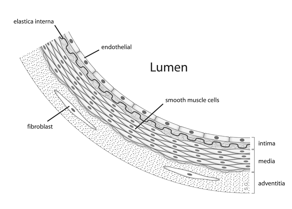